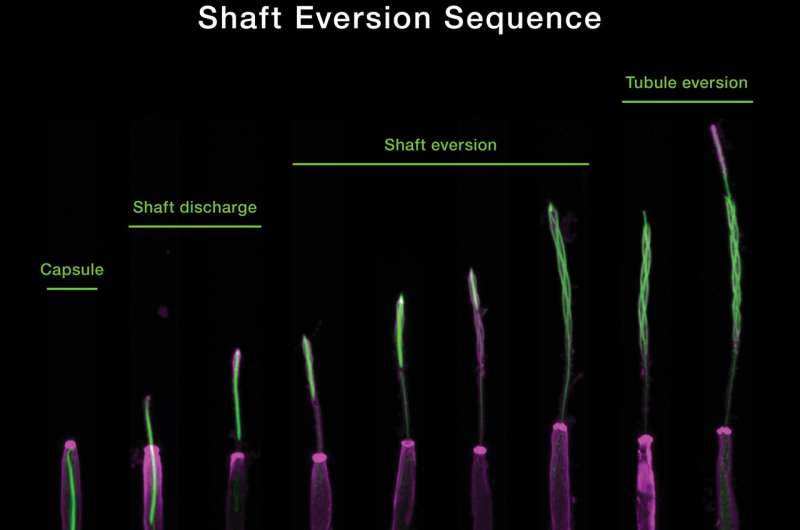
Fluorescent microscopy (prime) and mannequin (backside) present the mechanism for the ocean anemone’s stinging organelle in three distinct phases. Credit: Gibson Lab, Stowers Institute for Medical Research.
Summertime beachgoers are all too conversant in the painful actuality of a jellyfish sting. But how do the stinging cells of jellyfish and their coral and sea anemone cousins really work? New analysis from the Stowers Institute for Medical Research unveils a exact operational mannequin for the stinging organelle of the starlet sea anemone, Nematostella vectensis. The research, revealed on-line in Nature Communications on June 17, 2022, was led by Ahmet Karabulut, a predoctoral researcher within the lab of Matt Gibson, Ph.D. Their work concerned the appliance of cutting-edge microscopic imaging applied sciences together with the event of a biophysical mannequin to allow a complete understanding of a mechanism that has remained elusive for over a century. Insights from the work may result in useful functions in drugs, together with the event of microscopic therapeutic supply gadgets for people.
The Stowers group’s new mannequin for stinging cell perform supplies essential insights into the terribly complicated structure and firing mechanism of nematocysts, the technical identify for cnidarian stinging organelles. Karabulut and Gibson, in collaboration with scientists on the Stowers Institute Technology Centers, used superior imaging, three-dimensional electron microscopy, and gene knockdown approaches to find that the kinetic power required for piercing and poisoning a goal concerned each osmotic strain and elastic power saved inside a number of nematocyst sub-structures.
Serial pictures from electron microscopy had been used to create a 3D reconstruction of the ocean anemone’s stinging organelle. Credit: Gibson Lab, Stowers Institute for Medical Research.
“We utilized fluorescence microscopy, superior imaging strategies and 3D electron microscopy mixed with genetic perturbations to know the construction and working mechanism of nematocysts,” stated Karabulut.
Using these state-of-the-art strategies, the researchers characterised the explosive discharge and biomechanical transformation of N. vectensis nematocysts throughout firing, grouping this course of into three distinct phases. The first section is the preliminary, projectile-like discharge and goal penetration of a densely coiled thread from the nematocyst capsule. This course of is pushed by a change in osmotic strain from the sudden inflow of water and elastic stretching of the capsule. The second section marks the discharge and elongation of the thread’s shaft substructure which is additional propelled by the discharge of elastic power by means of a course of known as eversion—the mechanism the place the shaft turns inside out—forming a triple helical construction to encompass a fragile internal tubule adorned with barbs containing a cocktail of poisons. In the third section, the tubule then begins its personal eversion course of to elongate into the gentle tissue of the goal, releasing neurotoxins alongside the best way.
“Understanding this complicated stinging mechanism can have potential future functions for people,” stated Gibson. “This may result in the event of latest therapeutic or focused supply strategies of medicines in addition to the design of microscopic gadgets.”
During feeding, the ocean anemone’s tentacles seize brine shrimp. Credit: Gibson Lab, Stowers Institute for Medical Research.
The total stinging operation is accomplished inside only a few thousandths of a second, making it one of many quickest organic processes occurring in nature. “The earliest section of the firing of the nematocyst is extraordinarily quick and laborious to seize intimately,” stated Karabulut.
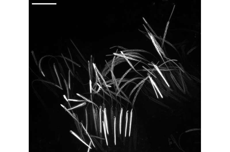
As is usually the case in basic organic analysis, the preliminary discovery was an accident of curiosity. Karabulut included fluorescent dye right into a sea anemone to see what it appeared like when the nematocyst-rich tentacles had been triggered. After making use of a mixture of options to each activate nematocyst discharge and concurrently protect their delicate substructures in time and house, he was shocked to have serendipitously captured a number of nematocysts at completely different levels of firing.
“Under the microscope, I noticed a surprising snapshot of discharging threads on a tentacle. It was like a fireworks present. I noticed nematocysts partially discharged their threads whereas the reagent I used concurrently and instantaneously mounted the samples,” stated Karabulut.

Sea anemone stingers at a number of levels of firing. Credit: Gibson Lab, Stowers Institute for Medical Research.
“I used to be capable of seize pictures that confirmed the geometric transformations of the thread throughout firing in a superbly orchestrated course of,” stated Karabulut. “After additional examination, we had been capable of absolutely comprehend the geometric transformations of the nematocyst thread throughout its operation.”
Elucidating the flowery choreography of nematocyst firing in a sea anemone has some attention-grabbing implications for the design of engineered microscopic gadgets, and this collaborative effort between the Gibson Lab and the Stowers Institute Technology Centers could have future functions for delivering medicines in people on the mobile degree.
Coauthors embody Melainia McClain, Boris Rubinstein, Ph.D., Keith Z. Sabin, Ph.D., and Sean McKinney, Ph.D.
Jellyfish venom capsule size affiliation with ache
More info:
Ahmet Karabulut et al, The structure and working mechanism of a cnidarian stinging organelle, Nature Communications (2022). DOI: 10.1038/s41467-022-31090-0
Provided by
Stowers Institute for Medical Research
Citation:
Inside the jellyfish’s sting: Exploring the micro-architecture of a mobile weapon (2022, June 23)
retrieved 23 June 2022
from https://phys.org/information/2022-06-jellyfish-exploring-micro-architecture-cellular-weapon.html
This doc is topic to copyright. Apart from any honest dealing for the aim of personal research or analysis, no
half could also be reproduced with out the written permission. The content material is supplied for info functions solely.


