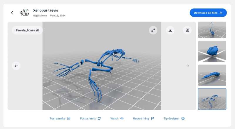
Discover the fascinating world of embryonic development and metamorphosis with the newly available 3D anatomical atlas of the African clawed frog, Xenopus laevis. This innovative tool provides researchers with valuable insights into the transformation process from tadpole to mature frog.
Previously, the lack of accessible data hindered the comprehensive understanding of these complex processes. To address this issue, the data has been converted into interactive 3D models using Sketchfab, allowing for easy viewing and exploration. Additionally, the files are available for 3D printing on Thingiverse, enhancing accessibility for science enthusiasts and educators.
Xenopus laevis has long been a key model organism in developmental biology, offering valuable insights into body plan reorganization during metamorphosis. However, the late developmental stages of this organism have been underrepresented in available datasets.
Lead scientist Dr. Jakub Harnos and his team from Masaryk University have bridged this gap by utilizing X-ray microtomography to create a detailed anatomical atlas of X. laevis. Through meticulous analysis of the 3D reconstructions, the researchers have identified crucial anatomical changes during the various developmental stages, shedding light on the intricate process of metamorphosis.
Explore the cutting-edge research and groundbreaking discoveries in the field of developmental biology with this invaluable resource. For more information, visit phys.org.


