


One grainy, gray-scale picture of a mind modified science and medication without end.
Half a century in the past, the primary CT picture of a affected person lifted the veil of invisibility that cloaks the inside of the human physique, offering scientists a window on our innards in contrast to any earlier than.
Today, docs within the United States alone order greater than 80 million scans per yr. X-ray computed tomography, or CT, is often the quickest means of getting a deal with on what’s inflicting a mysterious woe. CT scans can ferret out coronary heart illness, tumors, blood clots, fractures, inside bleeding and extra. The approach can provide surgeons a heads-up about what they’ll encounter inside a affected person, and information therapy for most cancers and different ailments.
“It answers so many questions quickly. That’s why it’s used,” says medical physicist Cynthia McCollough of the Mayo Clinic in Rochester, Minn.
A CT scan entails hundreds of X-ray measurements taken from a number of angles. Here’s the way it works: A supply of X-rays rotates across the physique, sending a beam of radiation via bone, blood and tissue, whereas rotating detectors measure the beam that makes it via.
Different supplies within the physique take in X-rays in another way. The calcium in bone vigorously sops up X-rays, for instance, whereas smooth tissues take in much less. So when the information collected by the detectors are stitched collectively by a pc, it may well type a cross-sectional view of what’s inside primarily based on the place X-rays are absorbed roughly. Moving the desk holding the affected person in order that the X-ray beam and detectors slide alongside the physique allows 3-D reconstructions of organs and different elements.
Sign Up For the Latest from Science News
Headlines and summaries of the most recent Science News articles, delivered to your inbox
Thank you for signing up!
There was an issue signing you up.
Over the years, scientists have regularly improved the know-how, making it sooner and better decision, and reducing the quantity of radiation that sufferers obtain. These improved CT scans have painted ever extra detailed landscapes of the human physique. It’s onerous to not marvel at the great thing about the inside world that the scans convey to the floor.
Here’s a glance again at among the main advances in CT scans over the past 50 years.
The CT scan is born
In 1971, radiologist James Ambrose at Atkinson Morley Hospital in London carried out the primary CT scan of a affected person in collaboration with engineer Godfrey Hounsfield, the know-how’s inventor. Hounsfield labored for the British electronics firm EMI, finest identified for its position because the file firm for the Beatles.
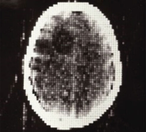 In 1971, the primary CT scan of a affected person (proven) laid naked the mind, uncovering a tumor (darkish circle at higher left).A. Maier et al/Medical Imaging Systems: An Introductory Guide 2018 (CC BY-ND 4.0)
The scan, a picture simply 80 pixels throughout and 80 pixels vast, didn’t present a number of element. But it showcased the know-how’s potential, revealing a tumor in a lady’s mind. A surgeon who later operated on the tumor is alleged to have marveled that “it looks exactly like the picture.”
Previously, if docs needed to search for a mind tumor, they injected air into the spinal column. Then they rotated the affected person to permit the air to bubble up into the realm across the mind to extend the distinction in customary X-ray pictures. The process “hurt like heck,” McCollough says. “Patients routinely vomited; it was like torture.” With CT scanning, that agony quickly turned a factor of the previous.
Any means you slice it
At first, CT scanners had been designed to picture solely the mind. But researchers shortly tailored the know-how to take cross-sectional pictures, or slices, at numerous factors alongside the physique. Then, within the early Nineteen Nineties, scanners had been created during which the X-ray supply scanned in a steady spiral across the physique, as a substitute of taking particular person cross-sections. That advance allowed an entire organ, such because the lungs, to be imaged in a single go.
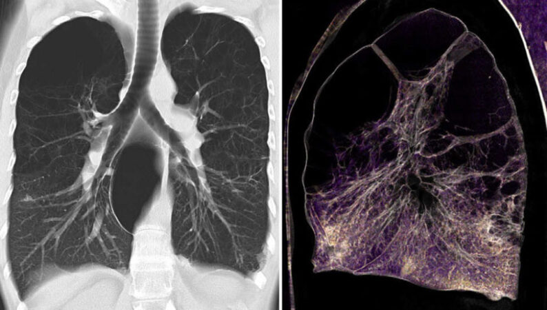 CT scanners with a number of rows of detectors can picture a number of cross sections, or slices, of the physique without delay, bettering picture decision. These pictures of lungs with emphysema had been taken with a 128-slice scanner, proven in a 2-D picture (left) and a colorized 3-D reconstruction (proper).P. Rogalla, C. Kloeters and P.A. Hein/Radiologic Clinics of North America 2009
But the picture decision alongside the size of the physique was nonetheless low. CT scanners with a number of rows of X-ray detectors solved this drawback by imaging a number of slices of the physique without delay alongside the spiral path.
CT scanner producers saved pushing up the variety of detectors, making scanners that would seize extra slices without delay. When detectors hit 64 slices within the early 2000s, McCollough says, “the real ‘Wow’ happened.” Scans may very well be quick, excessive decision and canopy a big size of the physique, suddenly. Today, scanners are much more subtle, utilizing as much as 320 slices.
Finally, the delicate complexity hidden contained in the human physique — from the intricate webs of blood vessels to the gracefully branching air passages of the lungs to the fragile but sturdy construction of bone — was there for all to see.
Doubling up
When McCollough’s father-in-law landed within the emergency room with a painful wrist, he languished for hours whereas docs tried to determine what was unsuitable. Eventually, McCollough remembers, she requested, “Can’t we send him over to CT?”
Thanks to a method known as dual-energy CT, docs had been capable of pinpoint the issue. Dual-energy CT, launched in 2006, makes use of two beams of X-rays at completely different energies, as a substitute of only one beam. Taking pictures this fashion permits the scanners to work out precisely what supplies are inside. Different supplies take in X-rays in several quantities, however with the intention to zero in on a particular kind of fabric, you wish to know the way that absorption modifications because the X-rays’ power modifications.
For instance, dual-energy CT can distinguish between several types of crystals that may type within the joints, inflicting arthritis. Urate crystals point out gout, and crystals containing calcium level to pseudogout. For McCollough’s father-in-law, the scan shortly uncovered the reason for his ache: pseudogout. In this fashion, CT scans can reveal the human physique at its most elementary stage, the supplies that make it up.
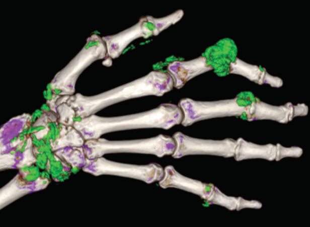 A dual-energy CT scan exhibits a telltale marker of gout, the presence of urate crystals (proven in inexperienced on this false-color 3-D illustration) in joints between the bones of the hand (white and purple).M.A. Desai et al/RadioGraphics 2011
Count on it
The X-ray detectors in CT scanners are the “secret sauce” of the know-how, McCollough says, as a result of the detectors are how the machines measure the X-rays within the first place. Most CT scanners measure that radiation not directly, first changing the X-rays into seen gentle after which changing that gentle into an electrical sign. A brand new period of CT know-how is reducing out the intermediary. In September, the U.S. Food and Drug Administration OK’d the primary “photon-counting” CT scanner.
X-rays are a sort of high-energy gentle, and like all gentle, they’re made up of particles known as photons. Photon-counting CT scanners measure particular person X-ray photons. The know-how additionally permits for crisper, extra detailed pictures and gives a measure of the energies of the photons, which, like dual-energy CT, identifies completely different supplies contained in the physique.
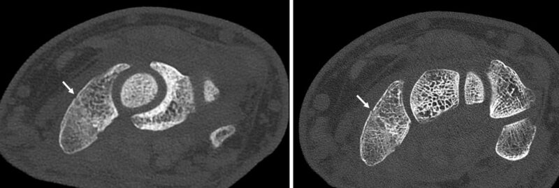 Photon-counting CT scans are prized for his or her high-resolution pictures. In a traditional CT scan of a damaged wrist (left, break indicated with an arrow), the construction of the bone is blurry in contrast with a photon-counting scan of the wrist (proper).Mayo Foundation for Medical Education and Research
Beyond the (human) physique
CT scans could have been invented for medication, however that didn’t cease different researchers from recognizing the know-how’s usefulness. Scientists in fields together with archaeology, biology and physics have harnessed the know-how to higher perceive all the things from mummified stays (SN: 8/20/20) to how cracks type in concrete to the anatomy of animals such because the earless monitor lizard (Lanthanotus borneensis).
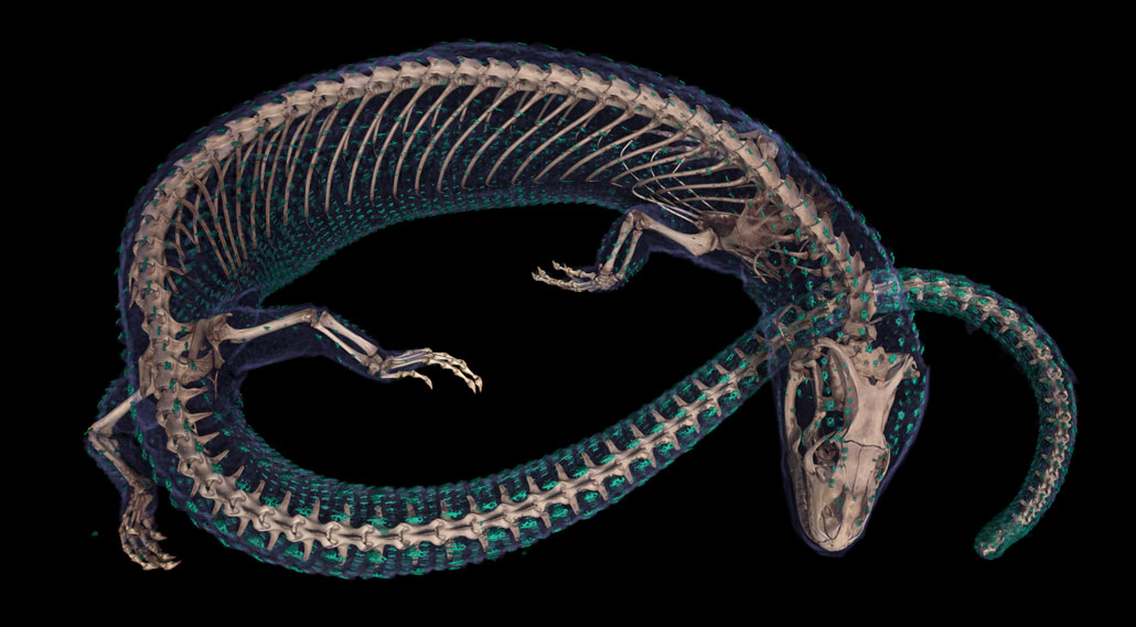 Created utilizing CT scanning, this colorized 3-D reconstruction of an earless monitor lizard reveals its skeleton (off-white) and bony armor (inexperienced).Edward Stanley/Florida Museum of Natural History/Univ. of Florida
This picture of L. borneensis comes from the Florida Museum of Natural History in Gainesville. Researchers there are making 20,000 CT datasets of preserved specimens that signify each vertebrate genus housed in U.S. pure historical past collections to disclose the animals’ inside anatomy. Anyone on this planet will be capable to entry these scans on-line.
The subsequent 50 years
Despite all of those advances, some researchers warning that physicians’ love affair with CT has gone too far, at the least within the United States, the place the scans are used extra typically than in lots of different international locations. Unneeded CT scans can have drawbacks: They could uncover unrelated findings that look regarding however are in all probability benign, which might result in costly and anxiety-inducing further testing. But foregoing CT scans may have damaging impacts, by slowing diagnoses for illnesses that resolve finest if handled shortly.
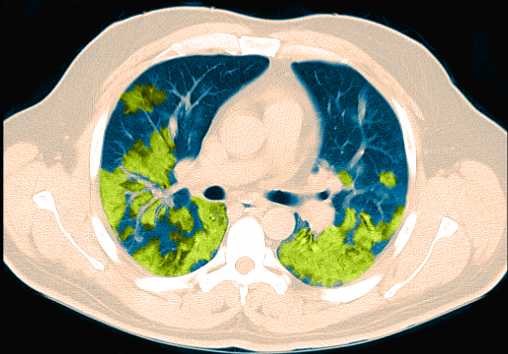 In sufferers with COVID-19, CT scans can reveal broken areas of the lungs, proven in inexperienced on this colorized picture of a cross part of the chest.Steven Needell/Science Source
There’s no denying that, when warranted, CT scans are a vital, lifesaving a part of the medical toolkit. Recently, the scans have been within the highlight for revealing injury achieved to the lungs by COVID-19 (SN: 4/27/20). The know-how appears poised to stay a mainstay of medication and science in future a long time, persevering with to scan its means into our hearts, lungs, brains and all over the place else.


