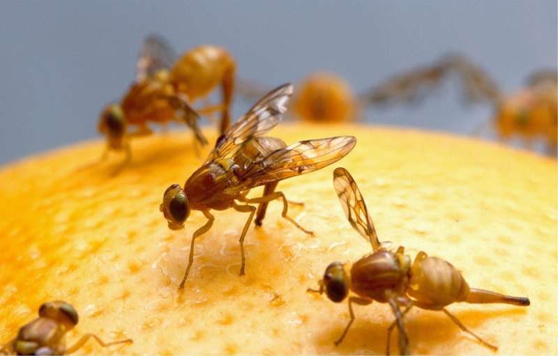
Credit: CC0 Public Domain
A groundbreaking achievement in neurobiological research has been made by a team of scientists supported by the National Institutes of Health (NIH)’s The BRAIN Initiative. Led by Davi Bock, Ph.D., Associate Professor of Neurological Sciences at UVM’s Robert Larner, M.D. College of Medicine, the team successfully mapped the entire brain of Drosophila melanogaster, commonly known as the fruit fly.
Published in Nature under the title “Whole-brain annotation and multi-connectome cell typing of Drosophila,” the study introduced a “consensus cell type atlas,” providing a comprehensive guide to understanding the various cell types in the fruit fly brain. With approximately 130,000 neurons, the fruit fly’s brain is significantly smaller than that of humans (86 billion neurons) or mice (100 million neurons).
The electron microscopy dataset used for creating this whole-brain connectome, known as FAFB (“Full Adult Fly Brain”), meticulously details every neuron’s shape and synaptic connections within the fly’s brain to identify and categorize all cell types present.
This detailed map will enable researchers to explore how different circuits collaborate to regulate behaviors such as motor control, courtship, decision-making, memory formation, learning processes, and navigation.
“To comprehend how brains function, we must understand how all neurons interact to facilitate cognitive processes,” noted study co-lead Gregory Jefferis, Ph.D.
“The functionality of most brains remains largely unknown. However with this complete wiring diagram for the fruit fly brain now available – a crucial step towards unraveling complex brain functions – other scientists have already begun using our shared data online to simulate how this tiny creature’s brain responds to its environment.”
2024-10-06 07:15:03
Post from phys.org
