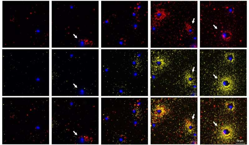
FluoroDOT assay photos of dendritic cells secreting protein 1 (TNFa, proven in crimson) and protein 2 (IL-6, proven in yellow) at completely different time factors (from left to proper: Unstimulated, stimulated for 30 minutes, 1 hour, 2 hours and three hours). The nuclei of the cell are proven in blue. The white arrows spotlight the cells that are both secreting just one protein or completely different quantities of the 2 proteins and completely different time factors. Credit: Srikanth Singamaneni/WashU
We have just lately witnessed the beautiful photos of distant galaxies revealed by the James Webb telescope, which have been beforehand seen solely as blurry spots. Washington University in St. Louis researchers have developed a novel methodology for visualizing the proteins secreted by cells with beautiful decision, making it the James Webb model for visualizing single cell protein secretion.
The researchers, led by Srikanth Singamaneni, the Lilyan & E. Lisle Hughes Professor of Mechanical Engineering & Materials Science within the McKelvey School of Engineering, and Anushree Seth, a former postdoctoral scholar in Singamaneni’s lab, developed the FluoroDOT assay, which they launched in a paper on August 5, 2022 within the journal Cell Reports Methods. The extremely delicate assay is ready to see and measure proteins secreted by a single cell in about half-hour.
In collaboration with researchers within the Washington University School of Medicine and different universities, they discovered that the FluoroDOT assay is flexible, low-cost and adaptable to any laboratory setting and has the potential to offer a extra complete take a look at these proteins than the broadly used current assays. Biomedical researchers look to those secreted proteins for info on cell-to-cell communication, cell signaling, activation and irritation, amongst different actions, however current strategies are restricted in sensitivity and might take as much as 24 hours to course of.
What makes the FluoroDOT assay completely different from current assays is that it makes use of a plasmonic-fluor, a plasmon-enhanced nanolabel developed in Singamaneni’s lab that’s 16,000 instances brighter than standard fluorescence labels and has a signal-to-noise ratio of almost 30 instances larger.
“Plasmonic-fluors are composed of metallic nanoparticles that function antenna to drag within the mild and improve the fluorescence emission of molecular fluorophores, thus making it an ultrabright nanoparticle,” Singamaneni mentioned.
This ultrabright emission of plasmonic-fluor permits the consumer to see extraordinarily small portions of secreted protein, which they’re unable to do in current assays, and measure the high-resolution alerts digitally utilizing the variety of particles, or dot sample, per cluster, or spot, utilizing a custom-built algorithm. In addition, it would not require particular gear. Singamaneni and his collaborators first printed their work with the plasmonic-fluor in Nature Biomedical Engineering in 2020.
The patent-pending plasmonic fluor know-how is licensed by the Office of Technology Management at Washington University in St. Louis to Auragent Bioscience LLC.
“Using a easy fluorescence microscope, we’re in a position to concurrently picture a cell together with the spatial distribution of the proteins secreted round it,” mentioned Seth, who labored on this mission as a postdoctoral scholar in Singamaneni’s lab and continues to work on it as a principal scientist (mobile purposes) for Auragent Bioscience. “We noticed fascinating secretion patterns for various cell sorts. This assay additionally permits concurrent visualization of two varieties of proteins from particular person cells. When the a number of cells are subjected to the identical stimuli, we will distinguish the cells which can be secreting two proteins on the identical time from those which can be solely secreting one protein or aren’t secreting in any respect.”
To validate the know-how, the workforce used proteins secreted from each human and mouse cells, together with immune cells contaminated with Mycobacterium tuberculosis.
One of the collaborators and co-authors, Jennifer A. Philips, MD, Ph.D., the Theodore and Bertha Bryan Professor within the departments of Medicine and Molecular Microbiology and co-director of the Division of Infectious Diseases within the School of Medicine, has used the FluoroDOT assay in her lab.
“When Mycobacterium tuberculosis infects immune cells, these cells reply by secreting essential immune proteins, referred to as cytokines,” Philips mentioned. “But not all cells reply to an infection the identical method. The FluoroDOT assay allowed us to see how particular person cells in a inhabitants reply to an infection—to see which cells are secreting and during which path. This was not doable with the older know-how.”
Team develops speedy SARS-CoV-2 take a look at primarily based on new plasmonic-fluor biolabeling know-how
More info:
Anushree Seth et al, High-resolution imaging of protein secretion on the single-cell stage utilizing plasmon-enhanced FluoroDOT assay, Cell Reports Methods (2022). DOI: 10.1016/j.crmeth.2022.100267
Provided by
Washington University in St. Louis
Citation:
‘Simple but {powerful}’: Seeing cell secretion like by no means earlier than (2022, August 5)
retrieved 5 August 2022
from https://phys.org/information/2022-08-simple-powerful-cell-secretion.html
This doc is topic to copyright. Apart from any honest dealing for the aim of personal research or analysis, no
half could also be reproduced with out the written permission. The content material is supplied for info functions solely.
