

Life is gorgeous in any respect scales, from massive to small. Sometimes that splendor is hid beneath literal scales.
A mesmerizing peek beneath the creating scales on the hand of an embryonic Madagascar large day gecko (Phelsuma grandis) gained first place within the 2022 Nikon Small World photomicrography competitors. The successful picture, stitched collectively from tons of of photographs taken over two days with a confocal microscope, was crafted by University of Geneva researchers Grigorii Timin and Michel Milinkovitch. The pair examine the genetics and physics of embryonic improvement.
The hand is artificially coloured to indicate budding nerves in cyan and constructions containing collagen in a variety of oranges and yellows. Collagen is a constructing block of life, says Milinkovitch. Knowing the place collagen is may help researchers higher perceive how our bodies and tissues develop.
Parts of bones which have began to calcify shine brightest within the picture, Timin says. Developing tendons and ligaments stretch as orange branches. Blood cells kind clusters or line up inside new blood vessels on the ideas of the gecko’s digits.
The picture highlights fantastic thing about all sizes, Milinkovitch says. The snapshot is “beautiful as a hand, you see this beautiful pattern of the fingers. Then you zoom, you see the spongy bones. And you zoom, you see the tendons. And you zoom, and you see the fibers that’s from the tendons. Then you zoom, and you see the blood cells.”
The gecko picture is considered one of 92 unimaginable photographs acknowledged on this 12 months’s competitors. The winners of the forty eighth annual contest have been introduced October 12. Here are a few of our different favorites.
Sign Up For the Latest from Science News
Headlines and summaries of the most recent Science News articles, delivered to your inbox
Thank you for signing up!
There was an issue signing you up.
Got milk?
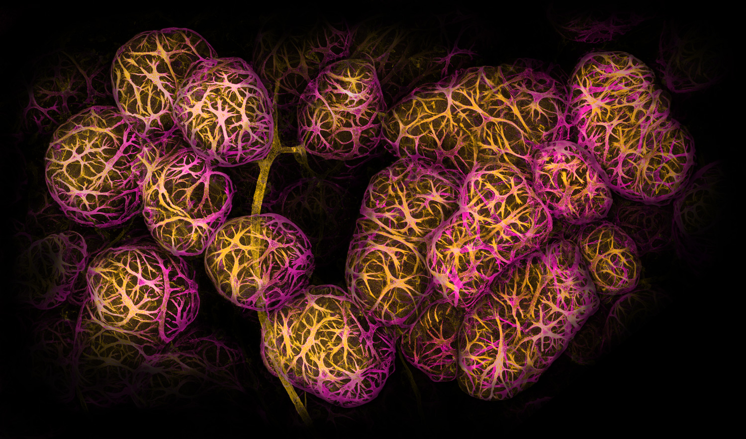

From a distance this {photograph} might appear to be a cluster of grapes. But every orb is a sprawling clump of cells inside breast tissue.
Cancer immunologist Caleb Dawson of the Walter and Eliza Hall Institute of Medical Research in Parkville, Australia, took 1000’s of photographs utilizing a confocal microscope to view tiny, musclelike cells that wrap round milk-producing spheres. He used dyes and antibodies to mark the cells yellow and magenta on this second-place-winning picture.
The cells reply to the hormone oxytocin, Dawson says. Oxytocin is launched throughout breastfeeding and helps squeeze milk out of the spheres, known as alveoli. Such photographs of lactating breast tissue may help researchers work out how immune cells maintain breast tissue and the infants it could possibly feed wholesome.
Snuffed candle
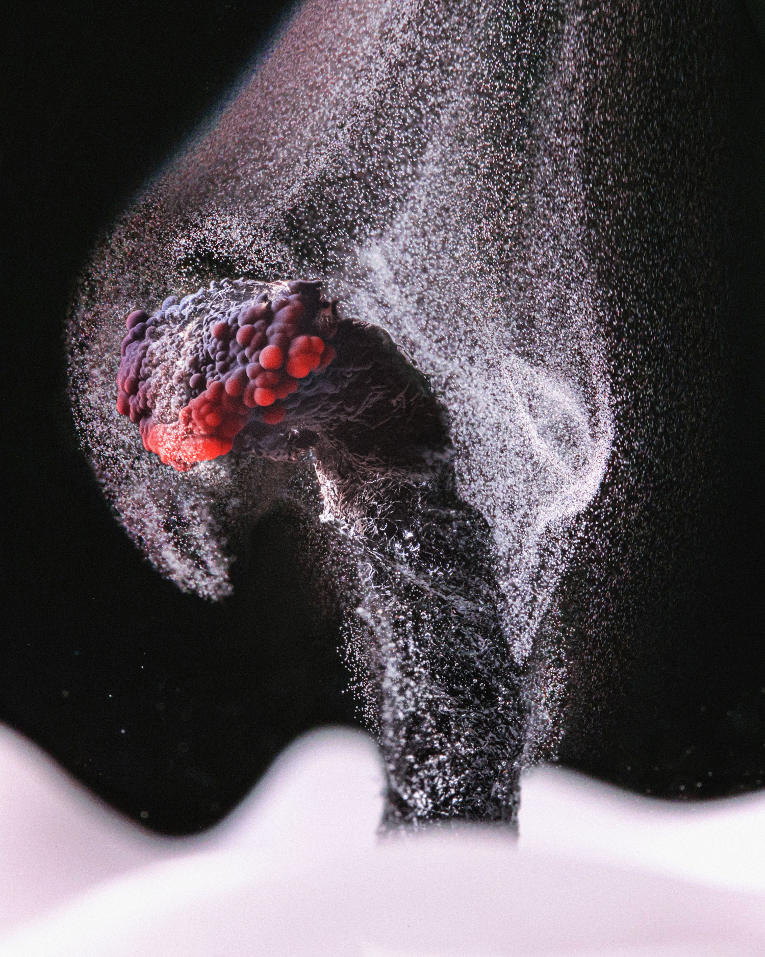

Ole Bielfeldt needed to be fast to seize the final gasp of an extinguished candle.
Candle wax is fabricated from hydrogen and carbon atoms, which flip largely into carbon dioxide when lit on hearth. But not all these hydrocarbons burn, as an alternative accumulating as soot on surfaces near the candle. “When the flame goes out, the glowing wick has enough heat left to break up the wax molecules for a while, but not enough to burn the carbon,” says Bielfeldt, a photographer in Cologne, Germany. “So you get a trail of smoke until it cools.”
Using a quick shutter pace and a vibrant LED mild, Bielfeldt managed to seize these unburned particles of carbon drifting away, incomes sixth place.
Iridescent slime mildew
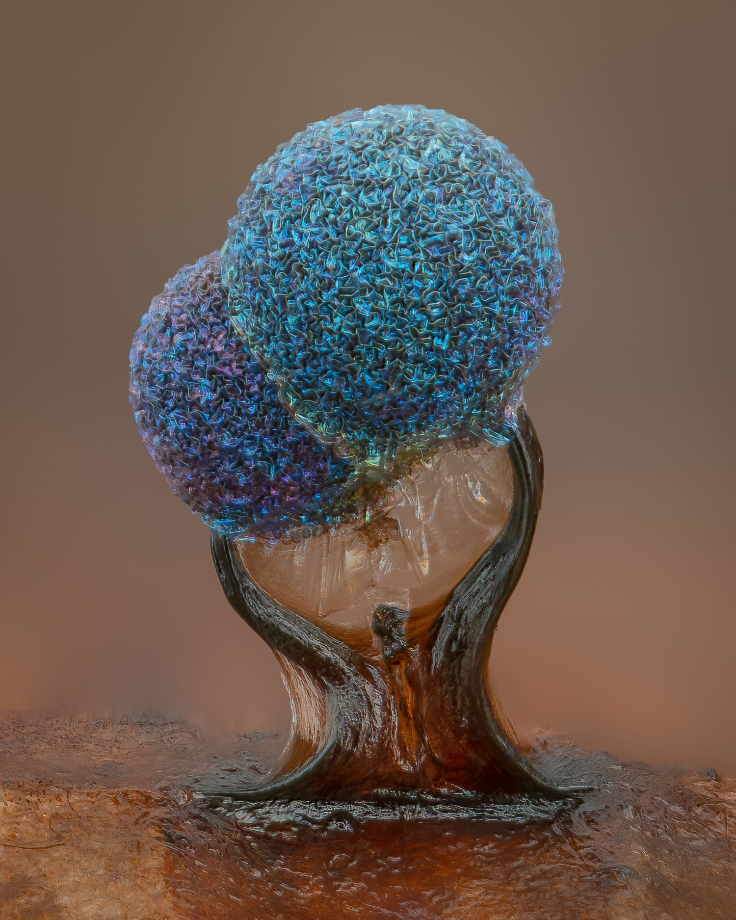
Hidden on leaves and decaying logs in moist forests are minuscule artworks like these Lamproderma slime molds.
In the dappled solar of an October day, photographer Alison Pollack of San Anselmo, Calif., noticed a glittery leaf as she was digging via a leaf pile. After taking the leaf dwelling and looking out via a microscope, she was transfixed by the crinkly heads and iridescence of slime mildew. Around 40 hours of labor and 147 mixed photographs later, Pollack had captured a putting snapshot that she likes to anthropomorphize as a nurturing relationship: dad or mum and youngster, two lovers, or brother and sister. The picture earned fifth place within the contest.
Most slime molds have clean heads, which launch spores into the atmosphere to breed. This pair might have dried too quick, stunting their improvement and leaving their heads wrinkly, Pollack says. But that’s OK, “because the texture to me is just gorgeous.”
A lethal predator
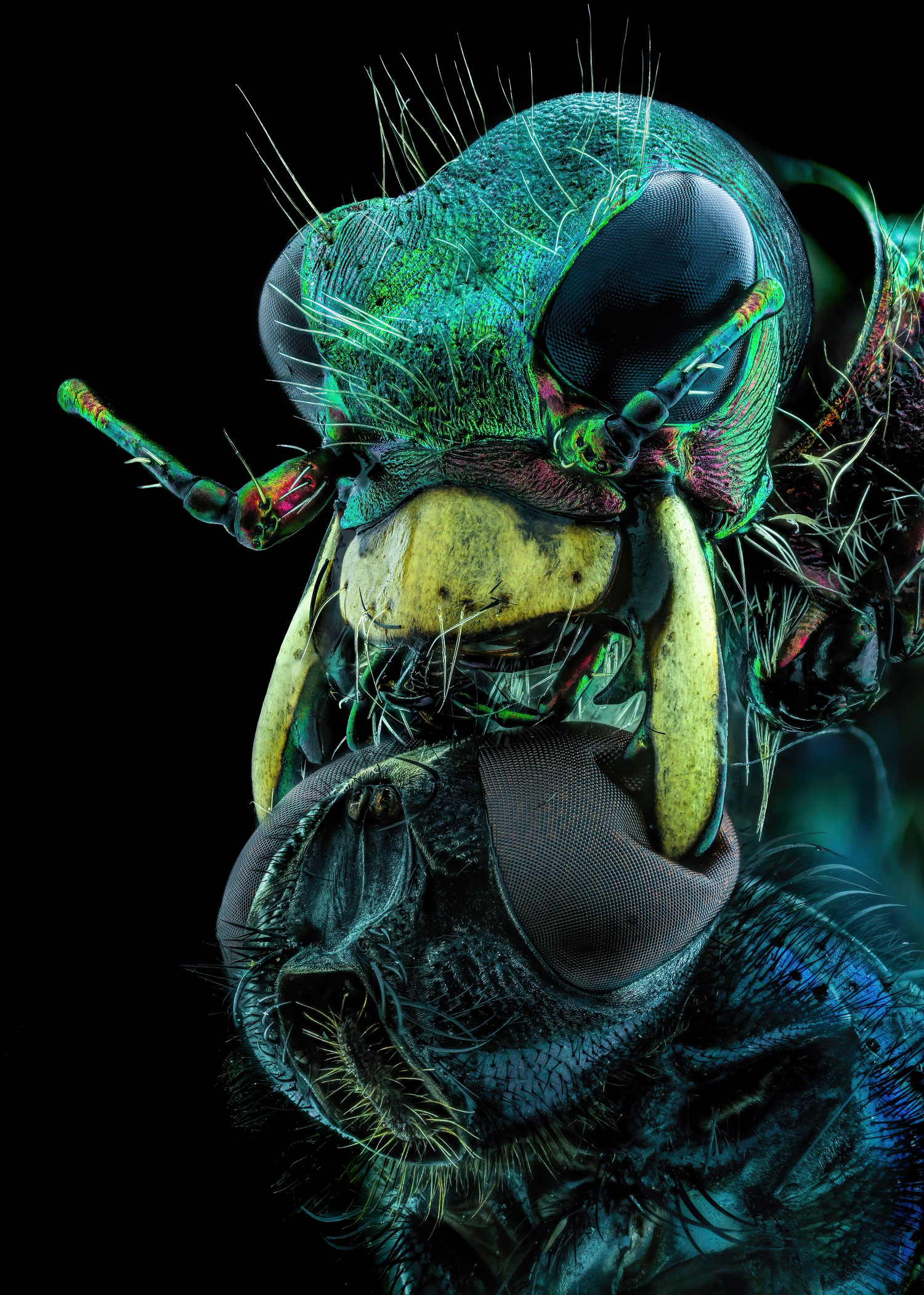

All concern the predacious tiger beetle, particularly this poor fly.
Murat Öztürk of Ankara, Turkey, nabbed tenth place on this 12 months’s competitors with an astounding — and unnerving — snapshot of a tiger beetle utilizing its mandibles to crush a fly by its eyes.
Tiger beetles (Cicindelinae) dash after their prey so rapidly that the bugs go quickly blind. The photographed beetle would have stopped a number of occasions to orient itself to determine the place the fly was, finally snatching its meal. Thanks to the beetle’s robust and sharp jaws, “the chances of survival of the creatures caught by this insect are very low,” Öztürk says.
Coral close-up
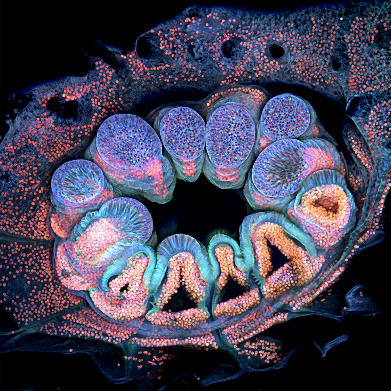

In Opal Reef off the coast of Australia, some cauliflower coral (Pocillopora verrucosa) polyps seem inexperienced. But the identical organism is remodeled when seen underneath a microscope within the lab.
To reveal the polyp’s particular person cells, marine scientist Brett Lewis of Queensland University of Technology in Brisbane, Australia, stitched collectively greater than 60 photographs taken over 36 hours. The coral naturally fluoresces a medley of blues, purples and pinks when uncovered to totally different wavelengths of sunshine. Algae residing contained in the polyp seem orange or pink, whereas the coral’s tissues shine blue. The picture gained twelfth place within the competitors.
One wonderful factor concerning the picture, Lewis says, is that in some areas, algal cells shine via a lightweight blue haze. That’s as a result of coral tissue is clear; algae give coral its shade.
Peeks of coral’s inner makings may help scientists perceive its biology, Lewis says. His work, as an example, goals to determine how younger polyps construct robust foundations after they connect to a floor — an necessary step in constructing or restoring coral reefs.